Motivation
Imaging Flow Cytometry is indispensable for cell analysis because images effectively provide certain messages about cells, such as cell size, shape, morphology, and distribution or location of biomarkers within cells. This wealth of information about a cell is crucial in various biological studies and medical diagnoses, such as early cancer detection.Importantly, Imaging Flow Cytometry is able to quantify cellular morphology and the intensity and location of fluorescent probes across the cell, on/in.
The data analysis performed on Imaging Flow Cytometry(IFC) image data contents still highly manual and subjective, i.e. using very limited image analysis techniques in combination with conventional flow cytometry gating strategies which are limited by the skill, experience and ingenuity of the data content analyst.
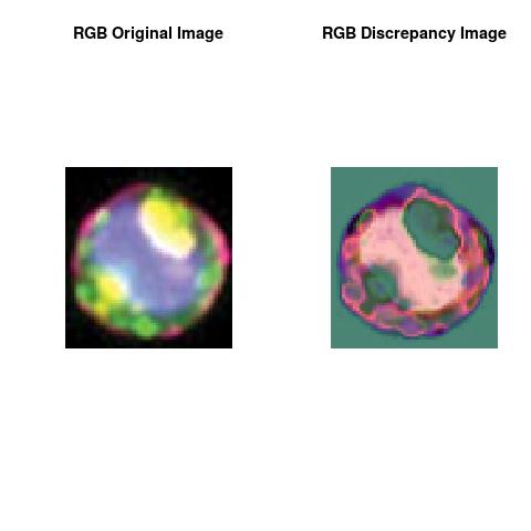
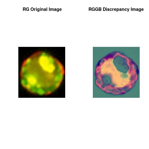
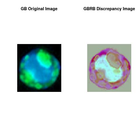
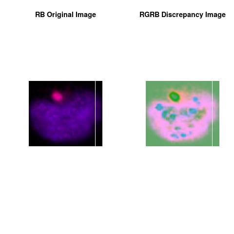
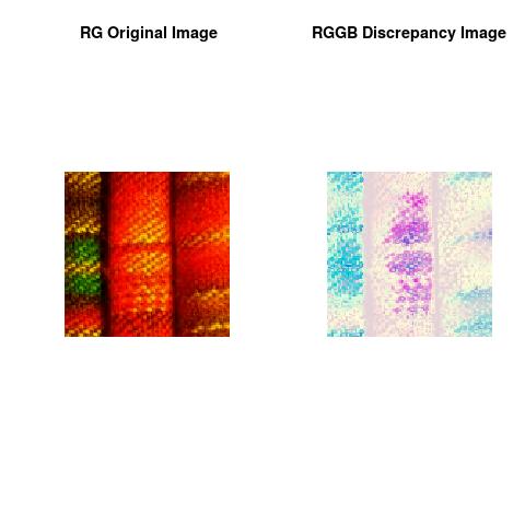
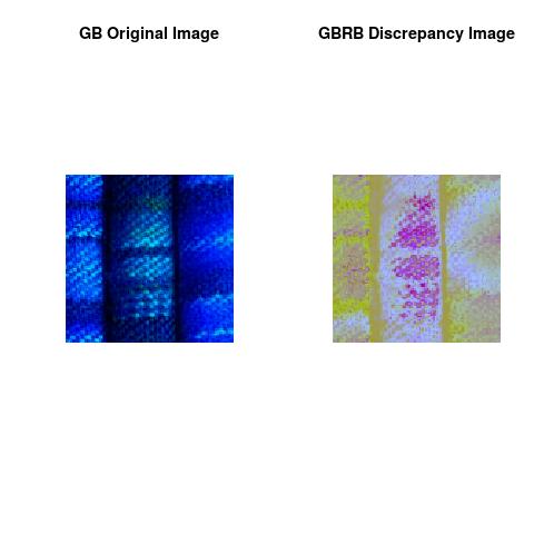
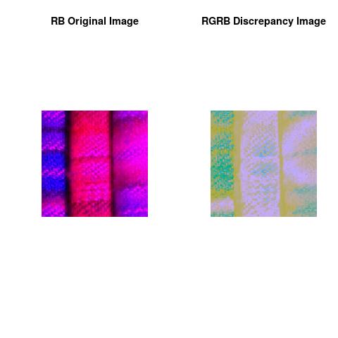
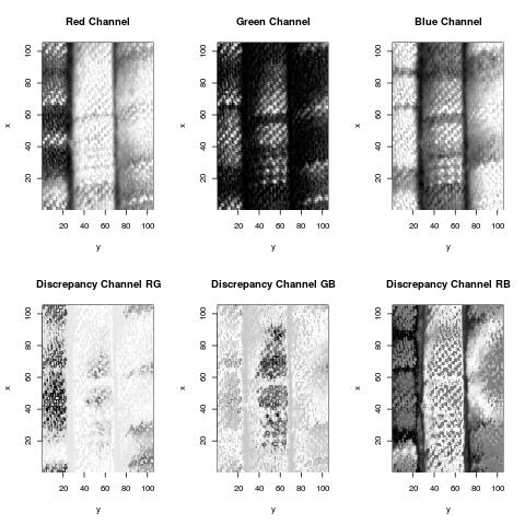
We are testing our recent developed software Fluorescence Imaging Analysis Using Deep Discrepancy Learning Process to automatically quantify the most informative and relevant features contained in the image.
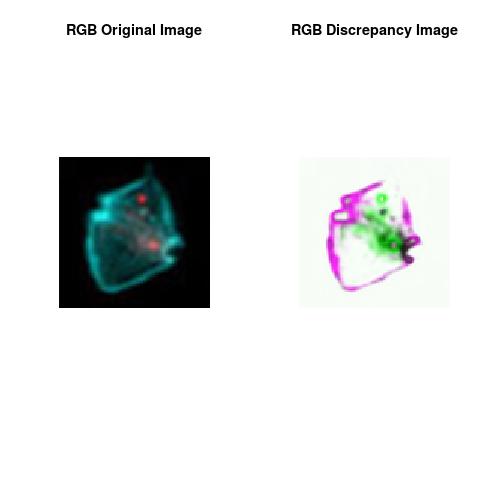
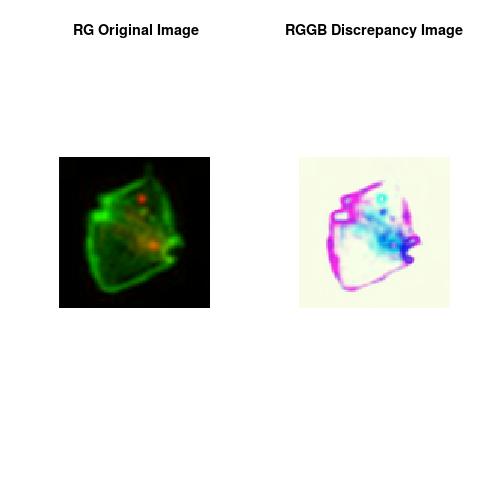
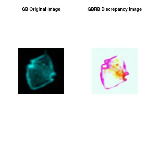
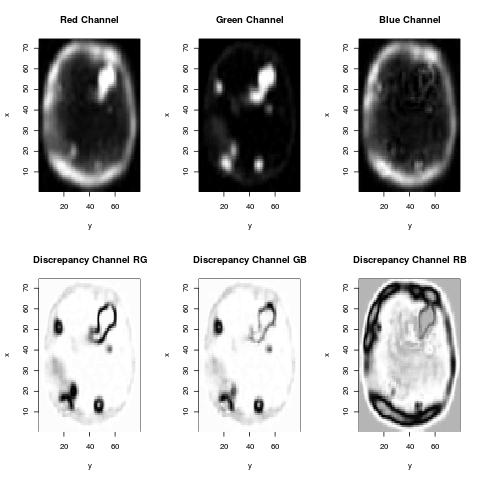
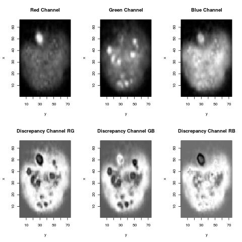
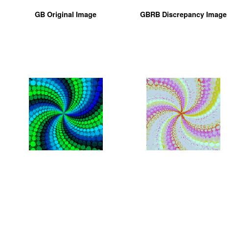
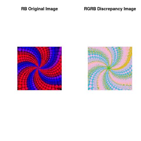
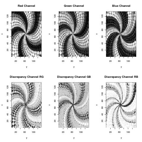
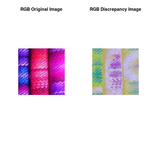
This approach is scalable and makes use of the totality or partially the provided channels in the images** and is simple to numerically implement without assuming any parametric form in the image data contents. The results showed that it captured the reflected pattern in the image with more insights about the colors spatial distribution where the pixel colors are either clumped or scarce.
**As some IFCs are Multi-spectral Imaging System.
Imaging Flow Cytometry and Discrepancy Image
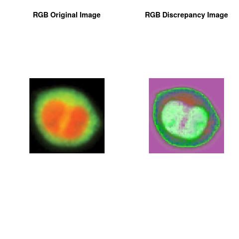
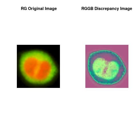
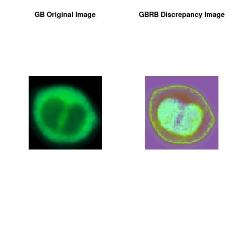
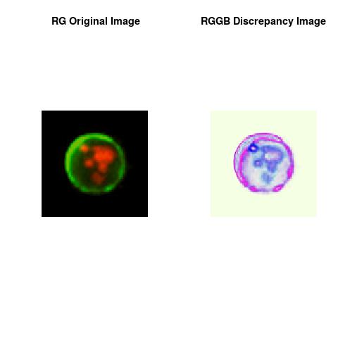
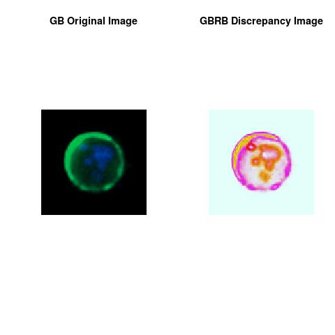
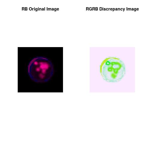
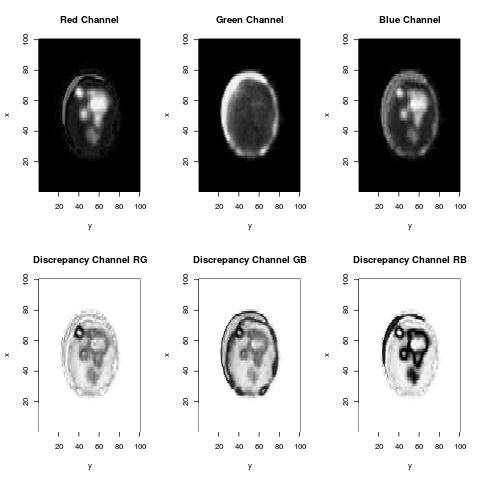
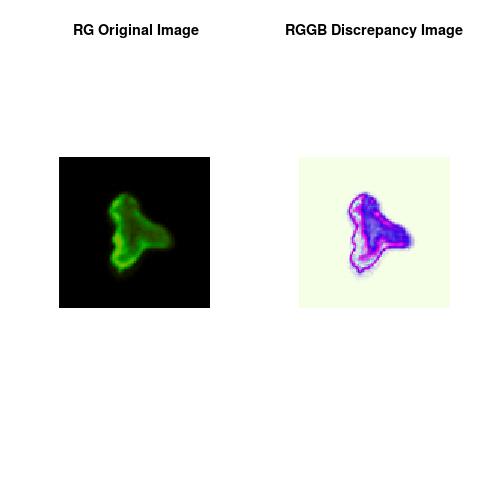
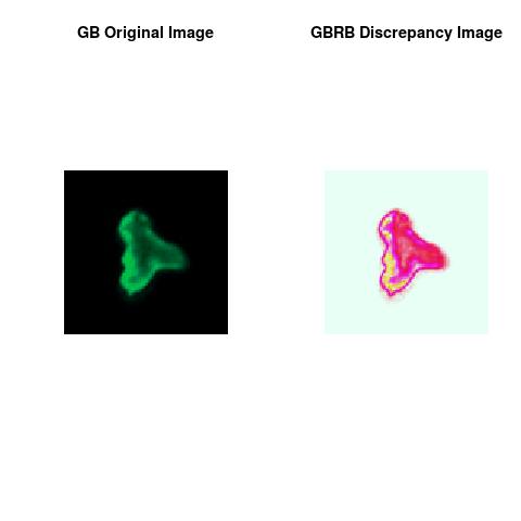
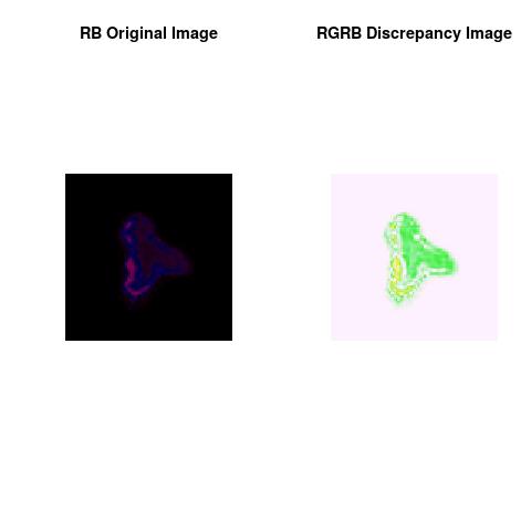
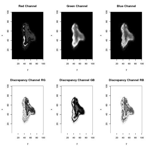
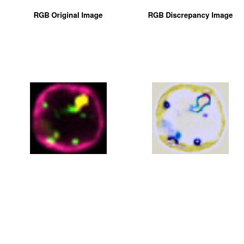
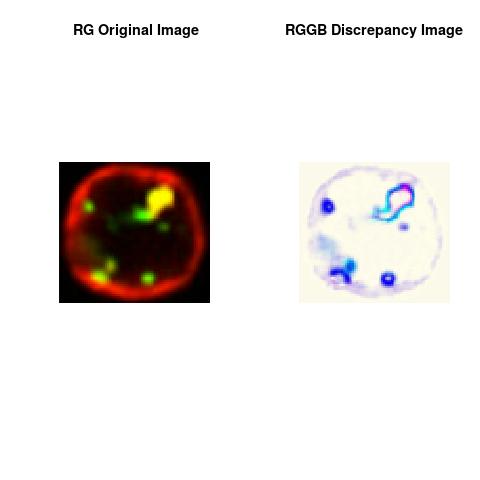
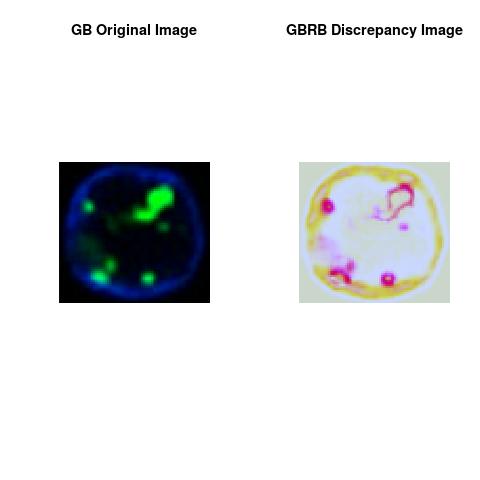
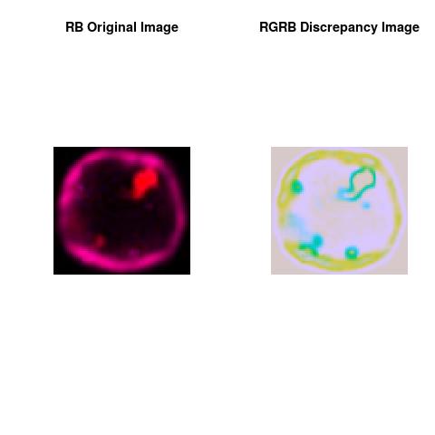
We are assessing the usefulness of the Discrepancy Images as companion images to a primary fluorescence image that provides further quantification (on measurement quality**) of the scarcity & abundance and distribution of the fluorescence contents in specified regions of interest(ROIs). **The relationship between sensitivity, resolution and IFC image quality.In Imaging Flow Cytometry, an Event can be a Pixel, Voxel or Multixel(as some IFCs are Multi-spectral Imaging System). Our illustration uses Pixel ( 2D image with 3 channels, Red-Green-Blue).
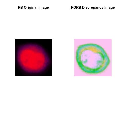
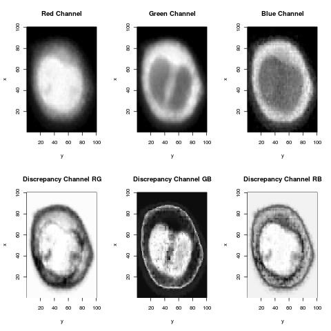
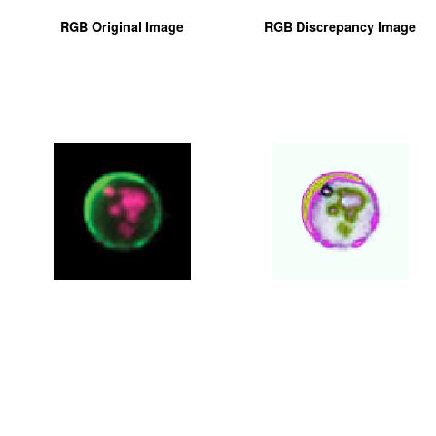
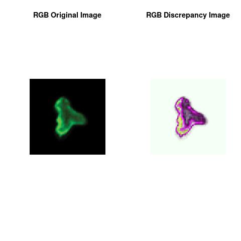
Figures show an application of Fluorescence Imaging Analysis Using Deep Discrepancy Learning Process to Imaging Flow Cytometry images from EMD Millipore Corporation/Amnis® Imaging Flow Cytometers.
Cell Signaling
( See Our Interpretation )
Molecular trans-location of transcription factors from the cytoplasm to the nucleus is a pivotal event in many processes critical to cellular activation, differentiation, and host defense. Existing methods used to measure nuclear trans-location have significant limitations.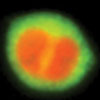
Cell-Death and Autophagy
( See Our Interpretation )
Cell death and survival, morphological characterization by microscopy remains the gold standard for accurately identifying the various types of cell death using characteristics such as nuclear condensation, nuclear fragmentation, membrane blebbing, cell shrinkage or swelling. In some cases cell death is preceded by an attempt at survival by autophagy and is identified by the clustering of the phagolysosome membrane-associated protein LC3.
Emerging Applications
Amnis® imaging flow cytometry platforms discriminate cells in flow objectively and statistically based on their appearance. This capability can therefore be used for any experiment that results in changes to the cell’s image.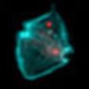
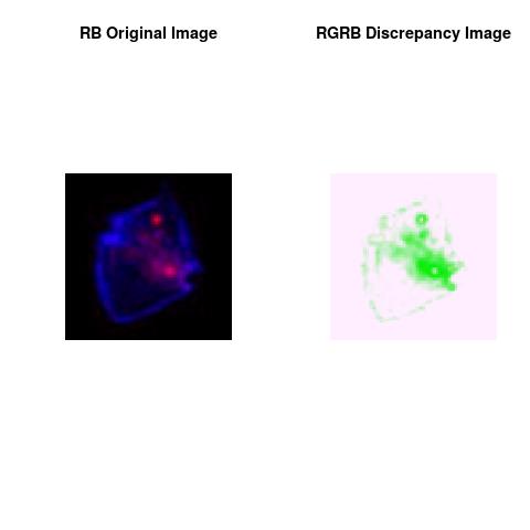
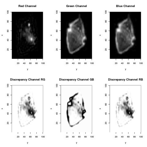
Morphology Analysis
( See Our Interpretation )
Distinguishing cells based on their morphologic differences is useful in the study of stem cell differentiation, hematology and oncology, and chemokine-induced shape change ...
Co-Localization
Microscopy is used to measure trafficking of molecules to sub-cellular compartments or the co-localization of proteins on, within, or between cells.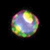
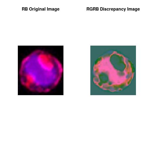
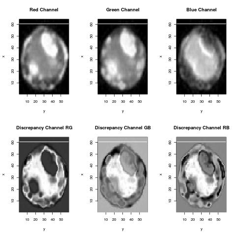
Internalization
Cellular processes such as drug uptake and processing by cells, entry of pathogens and toxins, or antigen processing and presentation are studied on Amnis® imaging flow cytometers by measuring internalization.
DNA Damage and Repair
The study of DNA repair mechanisms is important in the field of oncology where radiotherapy used to treat tumors induces double strand breaks (DSB). The DNA damage that occurs can be indirectly visualized with a microscope by immunostaining the repair proteins that are recruited to DSB foci.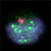
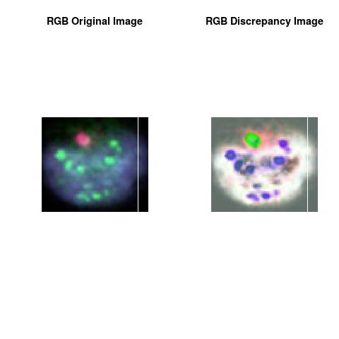
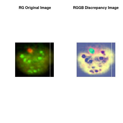
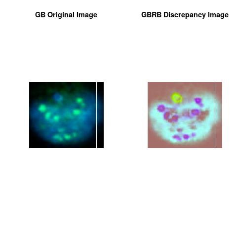
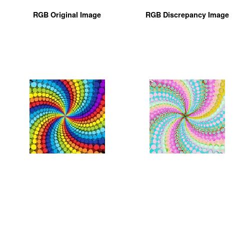
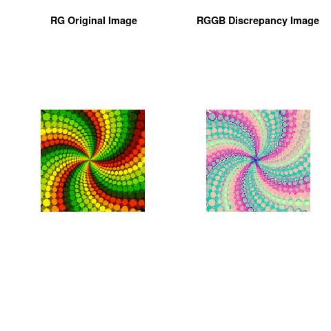
For an application to ordinary images, click this for more, Using Deep Discrepancy Learning Process.


Note
The theoretical part of this work was done when the author was in his PhD thesis (1994-1998), at the University of Savoie, Department of Mathematics, France. The work was shaped toward real applications accordingly to the learned scientific experience.This essay is mainly for Fluorescence Imaging Analysis and Discrepancy Images with application to Imaging Flow Cytometry (combining the speed and sample size of flow cytometry with the resolution and sensitivity of microscopy).
Author scientific profile:
Statistics and Applied Mathematics for Data Analytics, Identify opportunities to apply Mathematical Statistics, Numerical Methods, Machine Learning and Pattern Recognition to investigate and implement solutions to the field of Data Content Analytics. Data prediction via computational methods to predict from massive amounts of data (Big Data Content). These methods included clustering, regression, survival analysis, neural network, classification , ranking, deep discrepancy learning .
Author: Faysal.El.Khettabi@gmail.com , Living in Vancouver, BC, Canada.
The MIT License (MIT) Copyright 1994-2017, Faysal El Khettabi, Numerics&Analytics, All Rights Reserved.
The MIT License (MIT) Copyright 1994-2017, Faysal El Khettabi, Numerics&Analytics, All Rights Reserved.