Our Interpretation of IFC Using DDLP
The CCD Camera in Time-Delay Integration is able to empower the detection sensitivity to preserve the resolution and image quality in fast-moving objects. The cell is flowing as a 3D object, the instrument is able to adjust the speed of the cell with its internal velocity detection and as the cell is moving fast, the chance to spin during that short period of time is really minimal. Thus the instrument is able to map a three-dimensional object to a two-dimensional plane (the obtained image) by focusing on one side of the cell, as long as the cell does not spin, during its passage and the process of image acquisition, the image will be quasi-free from shutters. It's a quasi-"non-rotating" one-side 3D light source system for imaging multicolor-labeled cell.For instance, by using a dedicated marker to stain the membrane of the cell and a second marker to stain the nucleus. The instrument is able to construct an image of the cell as a 2D image where the areas(cytoplasm) between the membrane and nucleus are represented in an overlapping fashion.
The image may be obtained via forward detector or side scatter detector generated by the marker-cell-area activity with the light source. It's a similar system, the author is familiar with A three-dimensional x-ray scattering system for multi-parameter imaging of the human head.
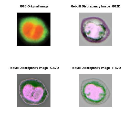
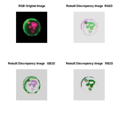
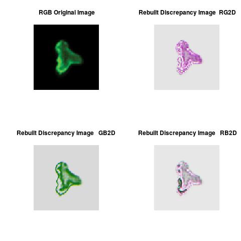
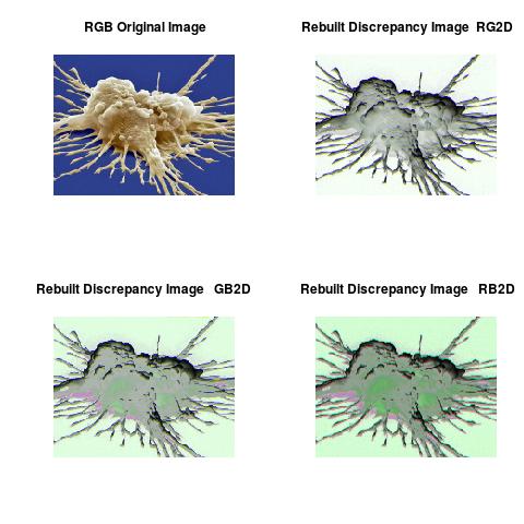
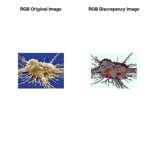
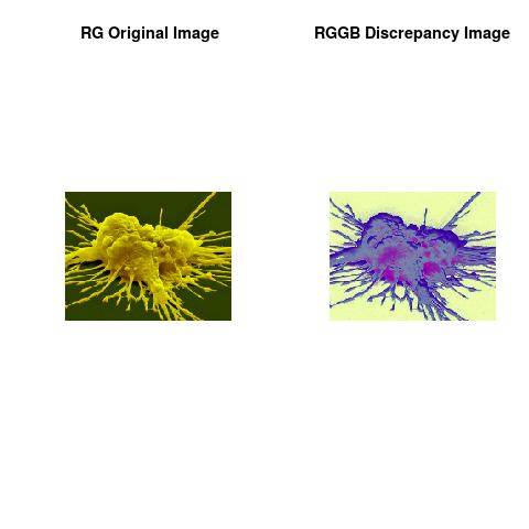
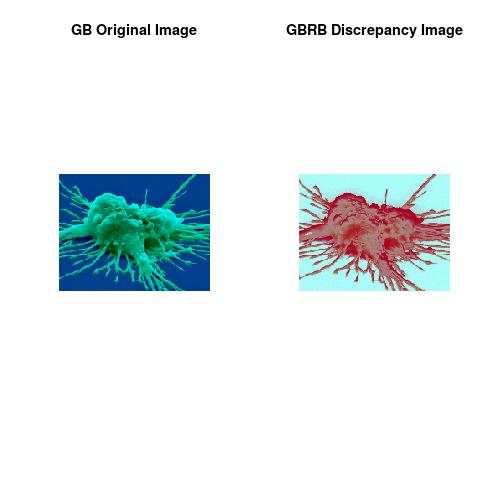
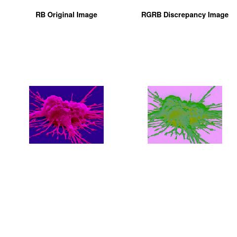
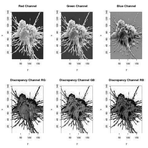
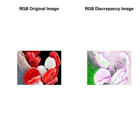
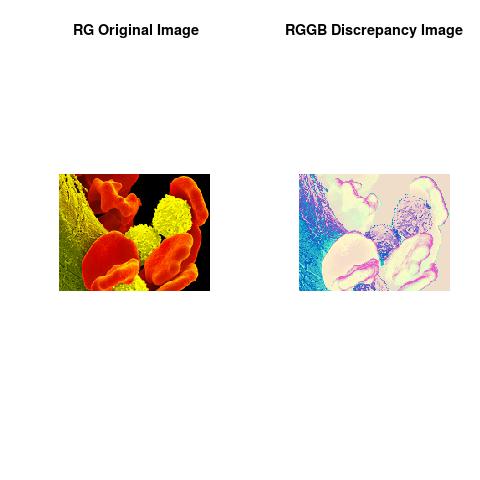
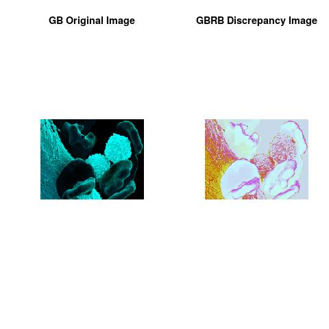
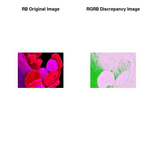
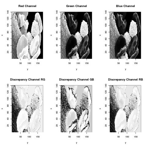
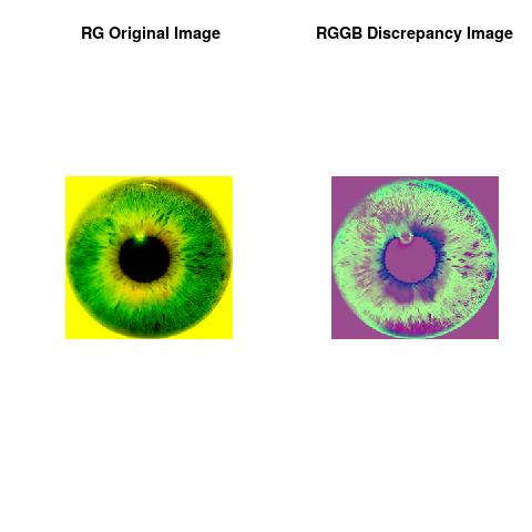
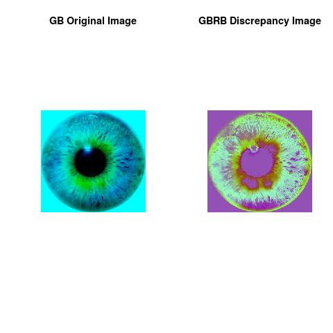
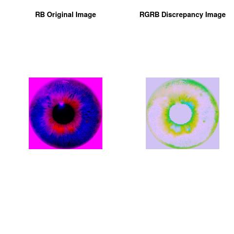
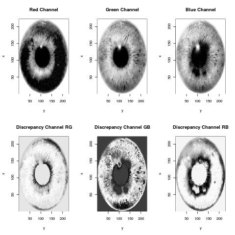
Our Deep Discrepancy Learning Process is typically used to locate objects and boundaries in the obtained image. More precisely, the process assigns to every pixel in the image a kind of score if the pixel shares certain characteristics or discrepancies, i.e. the pixel colors are either uniformly, clumped or scarce.
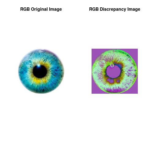
This framework was able to give insight about color distribution of the pixels and to allow access to many levels of information about image content. The results showed that the discrepancy framework captured the reflected pattern in the 2D image data and to provide a set of segments that collectively cover the entire image, or a set of contours extracted from the image, background, membrane, cytoplasm and nucleus.
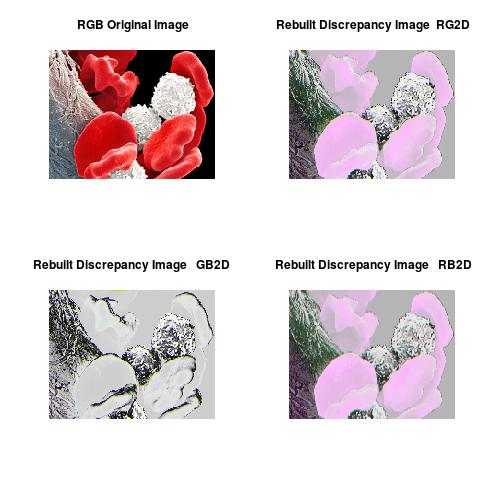
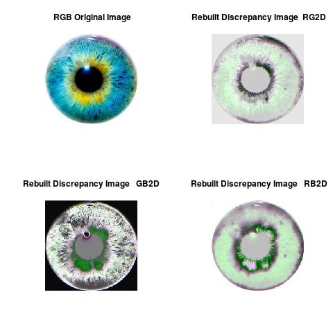
Each of the pixels in a region are similar with respect to some characteristic in the image data and show some discrepancies to other pixels, i.e. able to define adjacent regions. The discrepancy framework is an image segmentation of the primary image and is used to create 2D reconstruction of the flowing cell, see Figures with label "Rebuilt Discrepancy Image" as illustration.
Cell Signaling
Molecular trans-location of transcription factors from the cytoplasm to the nucleus is a pivotal event in many processes critical to cellular activation, differentiation, and host defense. Existing methods used to measure nuclear trans-location have significant limitations.


Cell-Death and Autophagy
Cell death and survival, morphological characterization by microscopy remains the gold standard for accurately identifying the various types of cell death using characteristics such as nuclear condensation, nuclear fragmentation, membrane blebbing, cell shrinkage or swelling. In some cases cell death is preceded by an attempt at survival by autophagy and is identified by the clustering of the phagolysosome membrane-associated protein LC3.


Morphology Analysis
Distinguishing cells based on their morphologic differences is useful in the study of stem cell differentiation, hematology and oncology, and chemokine-induced shape change ...


For an application to Fluorescence Imaging Analysis, click this for more, Using Deep Discrepancy Learning Process.
Note
The theoretical part of this work was done when the author was in his PhD thesis (1994-1998), at the University of Savoie, Department of Mathematics, France. The work was shaped toward real applications accordingly to the learned scientific experience.This essay is mainly for Fluorescence Imaging Analysis and Discrepancy Images with application to Imaging Flow Cytometry (combining the speed and sample size of flow cytometry with the resolution and sensitivity of microscopy).
Author scientific profile:
Statistics and Applied Mathematics for Data Analytics, Identify opportunities to apply Mathematical Statistics, Numerical Methods, Machine Learning and Pattern Recognition to investigate and implement solutions to the field of Data Content Analytics. Data prediction via computational methods to predict from massive amounts of data (Big Data Content). These methods included clustering, regression, survival analysis, neural network, classification , ranking, deep discrepancy learning .
Author: Faysal.El.Khettabi@gmail.com , Living in Vancouver, BC, Canada.
The MIT License (MIT) Copyright 1994-2017, Faysal El Khettabi, Numerics&Analytics, All Rights Reserved.
The MIT License (MIT) Copyright 1994-2017, Faysal El Khettabi, Numerics&Analytics, All Rights Reserved.